Giant Colony Morphologies
When I undertook this project I never imagined the microbiology involved and the hours I would spend in the lab just to get this project off the ground. Three months in the lab culturing organisms so far and I have found that just working with Brettanomyces spp. on various media agars can prove difficult. Each plate takes careful examination to ensure pure cultures are kept and maintained. Overtime this becomes easier and I have started to memorize traits each strain possess on the different media agars. The majority of the past few months have involved working with the Brettanomyces species cultured from Avery’s 15th Anniversary ale and cultures from White Labs yeast company. I have been culturing the Brettanomyces strains onto various medias listed previously and observed the morphologies and growth habits of each strain. The following is photos from the three White Labs Brettanomyces strains and their unique morphologies on various media agars. Along with culturing the different strains on various medias, careful observation under the microscope helps to observe strains general cell morphology but in no way gives a good indication to whether a pure culture is being observed due to the many growth phases and various cell shapes observed by a single culture at one time. I will try to post photos later and detail cell morphology under a microscope but that is too detailed to include in this post. For now here are some photos of the Brettanomyces spp. cultured on the various agar plates as this may be useful for any one trying to culture this incredibly fickle organism. The most difficult part of observing Brettanomyces is that little is written about the behavior and life cycle, if much is even known at all. I have discovered that even when a single colony is taken and streaked onto the agar plates not every resulting colony will grow exactly uniform or have exactly the same color or size. This could be due to a stage of the life cycle when streaked from a single colony or another phenomena, but it seems to be observed in nearly every strain.
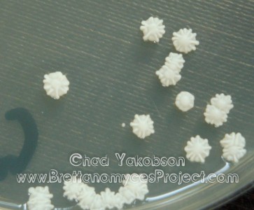
Brettanomyces bruxellensis (WLP650) on MYPG agar
Nearly all the colonies form a sand dollar
like pattern on top of the culture
with a round dome in the middle
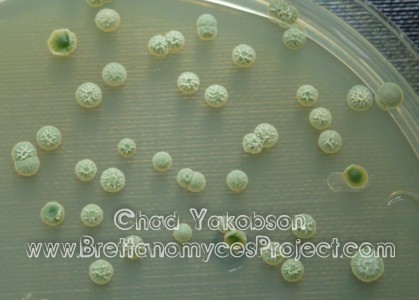
B. bruxellensis (WLP650) on WLN agar
Nearly all the colonies for a brain
like pattern on top of the culture
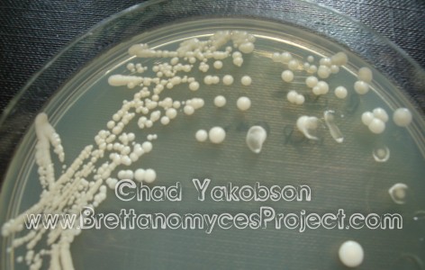
Brettanomyces claussenii (WLP645) grown on MYPG agar
White normal round colonies with a rather flat look
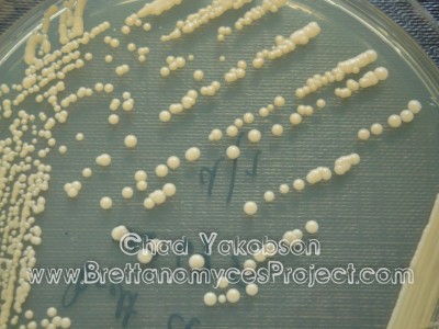
B. claussenii (WLP645) grown on MYPG+cycloheximide agar
A distinctive beige color is observable with the cycloheximide
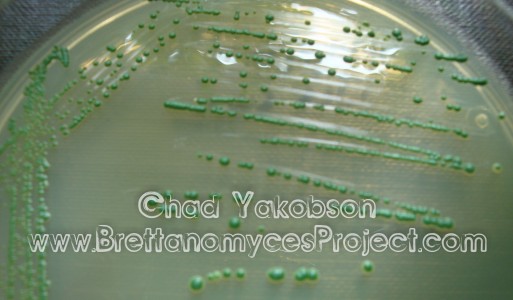
B. claussenii (WLP645) grown on WLN at 4 days since streaking
This species breaks the rules and does not
metabolize the Bromocresol Green
indicator as the literature states
New discovery, look how green the colonies remain
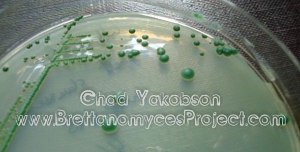
B. claussenii (WLP645) grown on
WLN at 7 days since streaking
Colonies still enlarging
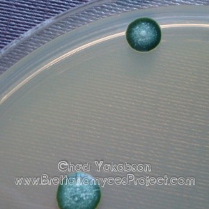
B. claussenii (WLP645) grown on
WLN at 10 days since streaking
Noticeable white dots appear on the top of the colony.
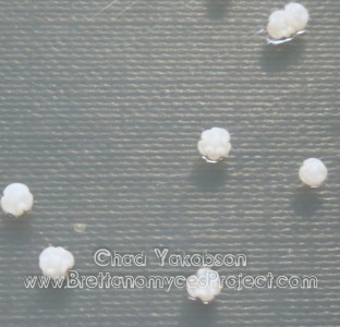
Brettanomyces lambicus (WLP653) on MYPG agar
colonies display a unique curved
ring like top structure
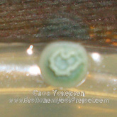
B. lambicus (WLP653) single colony
grown on WLN media with
a ring like top growth
on the colony
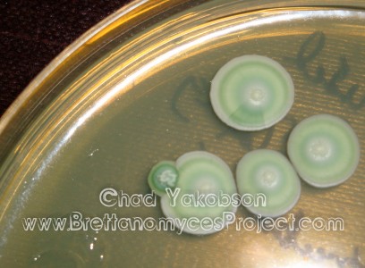
B. lambicus (WLP653) single colony
on WLN media growing into
the side of a Saccharomyces sp.
I have found Brettanomyces species like to grow on top of and within other yeasts.

I’m a biochemist at the University of California at San Francisco currently experimenting with Lambic-style brewing. I’ve been reading as much literature as I can find about the microbes used to brew lambic-style beer. Your website and research are awesome. Keep up the good work.
Have you been able to acquire any high resolution microscope images of the different strains you’re characterizing?
Just this week in the lab at the brewery we shot photos of all my strains along with two new ones which I’ve acquired. They are high res detailed photos of colony morphology on UBA and WLN media but not of the cells themselves under a microscope. I’m looking to get into a lab at the University where I’m now living to get those.. Will depend though, I would really like them as it would help for identification in brewing laboratories.