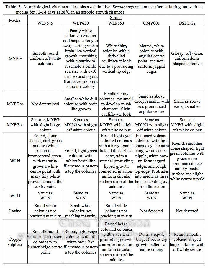
The morphologies of five Brettanomyces strains were observed to distinguish polymorphic variation on different media types and recorded for identification purposes (Table 2) see gallery for photos. B. bruxellensis (CMY001) showed the largest morphological variation when colony growths were compared between MYPG, WLN and CuSO4 medium, while B. bruxellensis (BSI-Drie) displayed similar morphological characteristics on each of the different plates. It was observed that both B. bruxellensis (WLP650) and B. lambicus (WLP653) formed distinctive colonies that produced a vertical, lip like growth on the top portion of the colonies. While the colonies of both strains had morphological differences they were the only two strains to form a vertical protruding growth on more than one media type. When grown on MYPG, B. claussenii (WLP645) formed round, off white, smooth colonies with no phenotypic variation observed. When grown on WLN medium, B. claussenii (WLP645) showed a large morphological change, the colonies were dark green and circular in shape with a dome like appearance. After 10-12 days, further growth in the form of a white center point occurred with many tiny white growths appearing around the center point. Minimal phenotypic variation was observed in the five strains. B. bruxellensis (WLP650) and B. bruxellensis (WY5112) were the exceptions with both showing phenotypic heterogeneity. Pearly white and beige colored colonies could be observed simultaneously on MYPG and UBA agars.
Throughout the study, eight strains of Brettanomyces were propagated in batch cultures and observed daily via microscope for pure culture verification. It was found that pure cultures could not accurately be confirmed by observing cell morphology under a microscope alone. Cell morphology was documented to change daily and the changes were not uniform between all strains. Given the varying ability of Brettanomyces yeast cells to either elongate or swell, cell size and morphology was found to be a highly variable characteristic within each strain. Some strains contained ogive, peanut shaped cells while others had the tendency to grow long and slender. Some strains swelled forming rugby ball like shapes and it wasn’t uncommon for a combination of morphologies to be viewed under the microscope all at once. Aguilar Uscanga et al. (2000) found changes in cell morphology were linked to the lack of nutrients when yeast extract was excluded from the growing medium and in the absence of ammonium sulfate. Since batch culture propagations were carried out in an all malt wort substrate during this study, changes in cell morphology due to a lack of nutrients should be ruled out and the probability of cell cycle dependent changes be considered. Further studies concentrating on the cellular morphology of Brettanomyces spp. could further the general understanding of the variances seen in pure cultures during growth phases.
For confirmation of pure cultures, batch culture propagations were routinely plated out onto MYPG, WLN and CuSO4 with giant colony morphologies observed. Each strain was found to form fairly stable, unique morphological characteristics on the different medias. Polymorphism was seen throughout the strains and was subject to the media used for culturing. Many of the strains formed detailed morphologies with unique characteristics not often observed in other genera of yeast, making them easy to distinguish. Uniformity amongst pure culture colonies was high with phenotypic heterogeneity only seen in two B. bruxellensis strains. Re-streaking of each colony type confirmed both colonies were from the same single strain and it is unknown why these two strains formed pearly white and beige colored colonies simultaneously on MYPG and UBA agar.

